
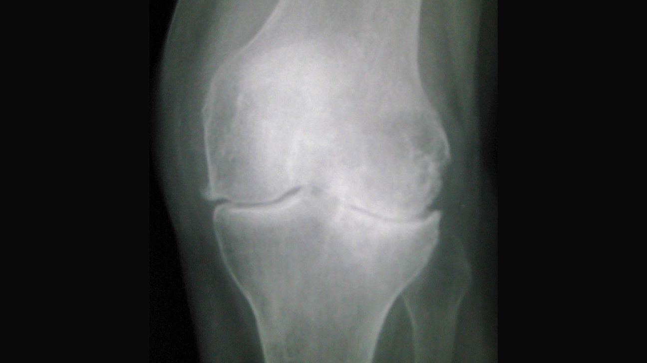
Doctors can use an X-ray (radiography) to view changes to your bones and their alignment in your knee joint. This can help them diagnose osteoarthritis.
If you’re experiencing unusual pain or stiffness in your knee joints, ask your doctor if osteoarthritis may be the cause. Your doctor may recommend an X-ray of your knee to find out.
This article explains everything you need to know about getting an X-ray to diagnose knee osteoarthritis.
X-ray imaging is a form of radiation that can pass through most objects, including the human body. During the test, the X-ray machine emits a brief burst of radiation that creates an image.
In this image, your bones appear white, your soft tissue appears gray, and the air appears black due to their different absorption levels.
X-rays are often the first-line imaging type for knee osteoarthritis. An orthopedist or bone doctor may refer you to get an x-ray to diagnose osteoarthritis after first examining you for signs or symptoms of osteoarthritis.
X-rays can’t capture images of cartilage. They can, however, reveal the loss of cartilage due to the narrowing of the space between bones, which is the most obvious symptom of osteoarthritis and other joint conditions in which cartilage has eroded. If your doctor wants to see more of the bone or soft tissue, they may also refer you to an MRI.
If the doctor diagnoses you with knee osteoarthritis, they will label your images based on severity grades 0 to 4 based on the
To get an X-ray of your knee, you’ll need to go to an X-ray imaging lab. There, a radiologist or an X-ray technician can take an X-ray and develop a detailed image of your bone structure for a better view of what might be affecting your joint area. You might also be able to have an X-ray done at your doctor’s office if it has X-ray equipment and a technician or radiologist on-site.
You don’t need to do much to prepare for an X-ray. Your radiologist may ask you to remove clothing covering your knees so that nothing blocks the X-rays from taking a fully detailed image.
If you’re wearing any metal objects, such as glasses or jewelry, your radiologist will likely ask you to remove them so they don’t appear on the X-ray image. Inform them of any metal implants or other metal objects in your body so that they know how to interpret the object on the X-ray.
If you’re a female assigned at birth (FAAB) of childbearing age, you may be asked if you are pregnant. If yes, your radiologist may not permit you to have an X-ray taken in order to keep the fetus safe. In this case, you might be able to have your knee examined with an ultrasound or other imaging technique.
Before the X-ray, the radiologist will take you to a small, private room. Others who may have come with you to the procedure may be asked to leave the room during the X-ray to protect them from radiation.
You’ll then be asked to stand, sit, or lie down in a position that allows the X-ray machine to capture the best possible image of your knee joint. You may feel slight discomfort depending on your position, but you’ll likely be given an object to lean or lie against, such as a pillow, to minimize your discomfort. You’ll also be given a lead apron to wear so that the rest of your body isn’t exposed to radiation from the X-rays.
Once you’re in position and have taken all the necessary precautions, you’ll be asked to stay still until the X-ray procedure is complete. You might be asked to hold your breath to make sure that you stay as still as possible. If you move during the X-ray, you may have to repeat the procedure more than once, as the image might be too blurry.
A simple joint X-ray shouldn’t take more than a few minutes, including any repeat procedures.
Exposure to some X-rays may increase the risk of developing cancer many years or even decades later after a test. However, the
Only young children may be noticeably sensitive to the radiation.
X-ray imaging results are usually available immediately after the procedure for you and your doctor to view. In some cases, your doctor may refer you to a specialist, such as a rheumatologist who specializes in arthritis, for further examination of your X-rays.
If your doctor doesn’t see any signs of cartilage loss or joint damage in your X-ray, your doctor may check the X-ray for signs of similar conditions, such as tendinitis or rheumatoid arthritis (RA).
Once a diagnosis is confirmed, your doctor will recommend the appropriate osteoarthritis treatment. They may also refer you to a physical or occupational therapist to help improve your knee’s flexibility.
What are the 4 features of knee osteoarthritis on X-ray?
The four tell-tale signs of osteoarthritis in the knee visible on an x-ray include joint space narrowing, bone spurs, irregularity on the surface of the joints, and sub-cortical cysts.
What is the best X-ray view for knee osteoarthritis?
X-rays are a standard way to get an image of your bone in order to identify signs of osteoarthritis.
They’re also preferred in many cases because they tend to be less expensive than MRIs, and the results are usually obtained more quickly. This allows your doctor to prescribe treatments or lifestyle changes that can decrease the constant pain and inflexibility that come with osteoarthritis.
In fact, while there are exceptions,